
Sleep is a bizarre state of consciousness. Perception almost completely disappears, we cease to feel our presence in space, and the passage of time undergoes strange disturbances. We experience dreams in which we don’t even remember who we are. And sometimes it seems to us that we fall into a black abyss, and our consciousness for several hours is close to non-existence.
The beginnings of sleep research
What happens inside our skulls when we sleep? The first answers came after the invention of the electroencephalograph. This device, through electrodes placed on the surface of the head, measures the weak electric field generated by the brain (this field is about 10,000 to 100,000 times weaker than that generated by a small 1.5 volt battery).
In the 1950s, Nathaniel Kleitman and Eugene Aserinsky demonstrated with an electroencephalograph that sleep is not a monolithic process, but is composed of alternating phases. Sleep has a distinct internal structure. During falling asleep, the frequency of brain waves gradually decreases and their amplitude increases, which constitutes the first phase of sleep. In the second phase, as the frequency of the waves continues to decrease, curious signals gradually appear, lasting from 0.5 to 2 s pulses with an internal frequency of 11 to 15 Hz [1]. These are called sleep spindles. They are generated by the thalamus, an internal brain structure that serves as a gateway to sensory impressions. Every few minutes, the spindles are preceded by strong, zigzag-like impulses called K-complexes, which are among the strongest electrical signals emitted by the brain. After that, the spindles fade away and the frequency of the brain waves decreases further. The sleeper enters the deepest phase of sleep. Brain waves now have a very low frequency: from about 0.5 to 2 Hz and a significant amplitude. These are called slow waves.
Then something unexpected happens. The brainwave frequencies quickly return to the range corresponding to the awake state, that is, the range from 15 to 60 Hz, but the sleeper does not wake up. Meanwhile, eyes begin to move much faster than in the previous phase. Hence, this phase of sleep was called REM (rapid eye movement), and the previous phase of sleep NREM (non-rapid eye movement).
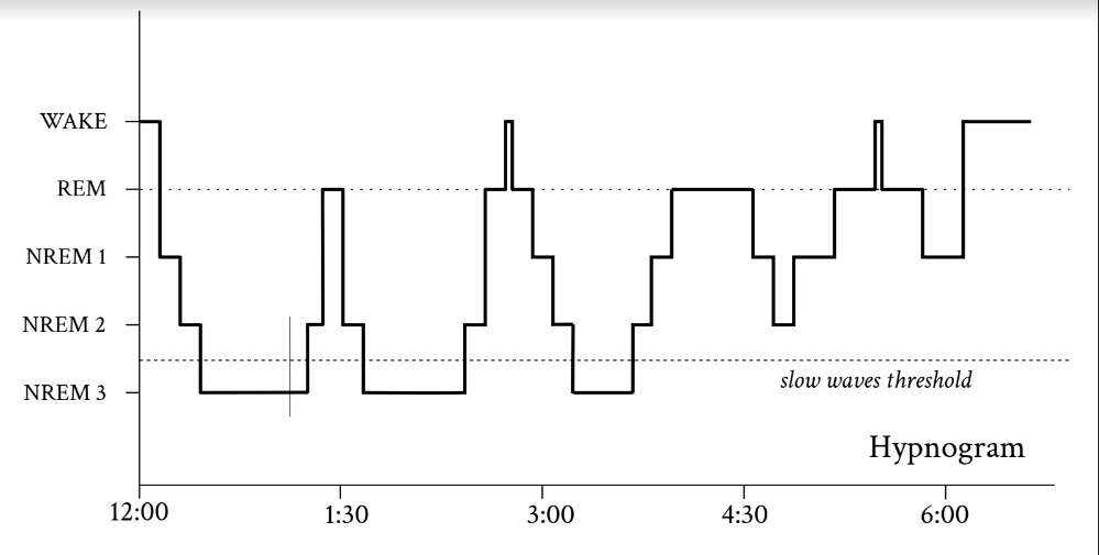
Figure 1 Example hypnogram showing transitions between sleep phases. The first part of sleep is dominated by deep sleep (NREM 3). REM sleep is more common in the second part. The hypnogram was created using the most common features of hypnograms, it does not represent a specific case.
The REM phase is where dreams often occur. However, we can also have dreams in the NREM phase, and their absence seems to be related to slow wave activity in the back of the brain [2]. When the mind travels into dream spaces during the REM phase, another surprising thing happens to our bodies. Muscles become completely flaccid because the brain loses control over them – we experience complete paralysis. This happens because the motor neurons of the brain that control the muscles become temporarily switched off. Or, more precisely, their electrical potential decreases. In this state, external impulses cannot stimulate the neurons enough to send signals to the muscles. Through paralysis during REM sleep, nature has protected us from performing the activities present in our dreams. This mechanism has a side effect – it sometimes happens that some sleepers wake up in the REM phase, but their bodies are still paralyzed. They cannot then make movements or speak, although their consciousness is already partially awake. This is often accompanied by a sensation of impotence, falling or being crushed. In this state, hallucinations sometimes occur. Fortunately, this state does not last long, passing most often after a few minutes.
After the first REM phase, we usually return to deeper sleep by starting another sequence of AWAKENESS/REM→ NREM → REM phases. This sequence is a sleep cycle and lasts approximately 90 minutes (±20 minutes). NREM deep sleep lasts longer in the first cycles, while REM sleep becomes longer in subsequent cycles.
Why does a sleep cycle exist? We don’t have a clear answer, but it is thought that the succession of phases is crucial to the learning process, i.e. tuning the strength of connections between neurons.
During the NREM phase, synaptic connections are normalized downward [3], thus removing unnecessary information noise enabling further learning. During the NREM [4] and REM [5] phases, the experiences of the day are also replayed in the hippocampus – the brain structure responsible for remembering. This means that the sequence of neuronal activation recorded during the day is repeated during sleep.
Understanding brain activity during sleep
Certainly, to better understand the functions of sleep, we should take a closer look at the phenomenon of slow waves in the NREM phase. What is the activity of neurons when the electroencephalograph records slow waves?
We can provide the answer by using another type of brain wave measurement: depth electrodes. Such electrodes are implanted in epilepsy patients to localize the focus of epilepsy. In addition, they provide valuable information about brain function. In particular, with their help we are able to record the discharges of individual neurons. These discharges, called action potentials, cannot be seen by the electroencephalograph.
It turns out that during the recording of slow brain waves, neuronal activity is strongly synchronized in a fairly regular rhythm. We can clearly discern periods in which neurons are active – the Up states – and periods of silence in which most neurons are inactive – the down states. The K-complex, which we mentioned earlier, is an electrical echo of a short, isolated lower state [6].
We also know where the source of the slow waves lies. Most of them originate in the frontal cortex, about 5 cm above the nasal root. The waves then propagate to the back of the head at about 3 m/s, and their amplitude decreases [7].
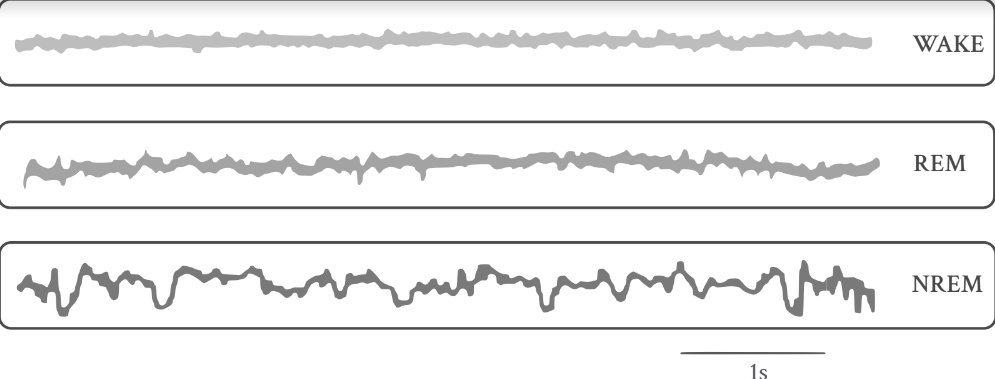
Figure 2 Electrical waves generated by the brain during: awakeness, REM sleep and NREM sleep. EEG waves recorded during rat sleep. [17]
What is the mechanism of Down and Up states? One hypothesis is that the Up state is the result of stimulation of the network by spontaneous local fluctuations in activity. Thanks to the dense number of neuronal connections in the human brain (on average, there are as many as 10,000 synapses per pyramidal neuron), the network is exponentially aroused to the Up state. Then, during activity in the neurons, a mechanism is triggered that naturally lowers the activity of the network – adaptation. In particular, potassium channels open in the cell membrane of neurons, as a result of which the electrical potential of neurons is lowered. At a certain point, it becomes impossible to maintain the activation of the entire network and the transition to the Down state occurs.
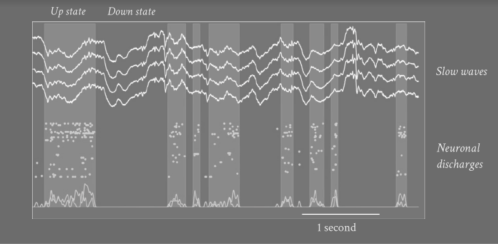
Figure 3 Slow waves and neuronal discharges recorded by a multi-electrode array implanted in an epilepsy patient. [18] The points correspond to the recorded action potentials of the neurons. We can clearly distinguish Upupper states with a lot of action potentials (lighter stripes) and Downlower states devoid of discharges (dark stripes).
With the knowledge of this mechanism, slow brain waves can be modeled in computational simulations. Using these, at the French NeuroPSI-CNRS (Centre national de la recherche scientifique), our research team was able to explain the statistical differences between the slow waves occurring in deep sleep and those generated during anesthesia [8]. Central to these differences, as well as to the process of slow wave generation itself, is the concentration of neuromodulators such as acetylcholine, serotonin and norepinephrine.
Slow waves and the sleep spindles present in the NREM-2 phase are crucial in the process of preservation of memory traces, which we call consolidation. It is presumed that it is during slow-wave sleep that information is transferred from short-term memory to long-term memory [9]. A decrease in the length of slow-wave sleep has been shown to be correlated with a weaker ability to remember [10]. Unfortunately, in the aging process, the length of slow-wave sleep decreases at a rate of about 2% per decade [11], thus reducing memory abilities.
Application of the research
Understanding the mechanism of slow-wave may allow the development of methods of deep sleep prolongation.. This is particularly important for improving sleep quality in the elderly and patients with sleep disorders. There are already first techniques for enhancing slow waves, such as auditory stimulation [12], slow-wave electric field stimulation (so-tDCS) [13], or pharmacological methods (interleukin-6 [14] and sodium oximebutyrate [15]). These have shown some effectiveness in improving sleep quality and intellectual performance [16].
However, even without them, we can improve the quality of our sleep starting with the simplest methods – for example, by limiting caffeine and alcohol, which is known to strongly disrupt the REM phase. Reducing evening exposure to blue light, which disrupts melatonin production, also helps improve sleep quality.
Editorial Board’s Note: Certainly, highlighting the application of these techniques can have a beneficial impact on the mental and physical health of Poles, potentially increasing the efficiency and quality of both their social and professional lives. This would undoubtedly have a positive influence on the Polish market.
References:
- Purcell, S., Manoach, D., Demanuele, C. et al. Characterizing sleep spindles in 11,630 individuals from the National Sleep Research Resource. Nat Commun 8, 15930 (2017).
- Siclari, F., Baird, B., Perogamvros, L. et al. The neural correlates of dreaming. Nat Neurosci 20, 872–878 (2017)
- Tononi G, Cirelli C. Sleep function and synaptic homeostasis. Sleep Med Rev. 2006 Feb;10(1):49-62.
- Lee AK, Wilson MA. Memory of sequential experience in the hippocampus during slow wave sleep. Neuron. 2002;36(6):1183–1194.
- Louie K, Wilson MA. Temporally structured replay of awake hippocampal ensemble activity during rapid eye movement sleep. Neuron. 2001;29(1)
- Cash SS, Halgren E, Dehghani N, Rossetti AO, Thesen T, Wang C, Devinsky O, Kuzniecky R, Doyle W, Madsen JR, Bromfield E, Eross L, Halász P, Karmos G, Csercsa R, Wittner L, Ulbert I. The human K-complex represents an isolated cortical down-state. Science. 2009 May 22;324(5930):1084-7.
- Hangya B, Tihanyi BT, Entz L, Fabó D, Erőss L, Wittner L, Jakus R, Varga V, Freund TF, Ulbert I. Complex propagation patterns characterize human cortical activity during slow-wave sleep. J Neurosci. 2011 Jun 15;31(24):8770-9.
- Nghiem TE, Tort-Colet N, Górski T, Ferrari U, Moghimyfiroozabad S, Goldman JS, Teleńczuk B, Capone C, Bal T, di Volo M, Destexhe A. Cholinergic Switch between Two Types of Slow Waves in Cerebral Cortex. Cereb Cortex. 2020 May 18;30(6):3451-3466
- Diekelmann, S., Born, J. Slow-wave sleep takes the leading role in memory reorganization. Nat Rev Neurosci 11, 218 (2010)
- Ferrarelli F, Kaskie R, Laxminarayan S, Ramakrishnan S, Reifman J, Germain A. An increase in sleep slow waves predicts better working memory performance in healthy individuals. Neuroimage. 2019 May 1;191:1-9; Bartsch, U., Simpkin, A.J., Demanuele, C. et al. Distributed slow-wave dynamics during sleep predict memory consolidation and its impairment in schizophrenia. npj Schizophr 5, 18 (2019)
- Zolovska, J.P. Shatkin, in Encyclopedia of Sleep, 2013
- Ngo HV, Martinetz T, Born J, Mölle M. Auditory Closed-Loop Stimulation of the Sleep Slow Oscillation Enhances Memory. Neuron. 2013;78(3):545–53.
- Antonenko D, Diekelmann S, Olsen C, Born J, Mölle M. Napping to renew learning capacity: enhanced encoding after stimulation of sleep slow oscillations. Eur J Neurosci. 2013. October;37(7):1142–51; Göder R, Baier PC, Beith B, Baecker C, Seeck-Hirschner M, Junghanns K, et al. Effects of transcranial direct current stimulation during sleep on memory performance in patients with schizophrenia. Schizophr Res. 2013;144(1-3):153–4.
- Benedict C, Scheller J, Rose-John S, Born J, Marshall L. Enhancing influence of intranasal interleukin-6 on slow-wave activity and memory consolidation during sleep. FASEB J. 2009 Oct;23(10)
- Walsh JK, Hall-Porter JM, Griffin KS, Dodson ER, Forst EH, Curry DT, Eisenstein RD, Schweitzer PK. Enhancing slow wave sleep with sodium oxybate reduces the behavioral and physiological impact of sleep loss. Sleep. 2010 Sep;33(9):1217-25.
- Can Slow-Wave Sleep Enhancement Improve Memory? A Review of Current Approaches and Cognitive Outcomes Yujie Zhang 1, Reut Gruber 2
- Alejandra Mondino et al., Power and Coherence in the EEG of the Rat: Impact of Behavioral States, Cortical Area, Lateralization and Light/Dark Phases (2020). Creative Commons 4.0.
- Trang-Anh E Nghiem, Núria Tort-Colet, Tomasz Górski, Ulisse Ferrari, Shayan Moghimyfiroozabad, Jennifer S Goldman, Bartosz Teleńczuk, Cristiano Capone, Thierry Bal, Matteo di Volo, Alain Destexhe, Cholinergic Switch between Two Types of Slow Waves in Cerebral Cortex, Cerebral Cortex, bhz320
Dr Tomasz Gorski is a neuroscientist specializing in computational neuroscience. After obtaining his PhD in theoretical physics at the University of Lodz, he worked at research centers in Poland, France and Switzerland. ©Tomasz Górski
Tomasz Górski
