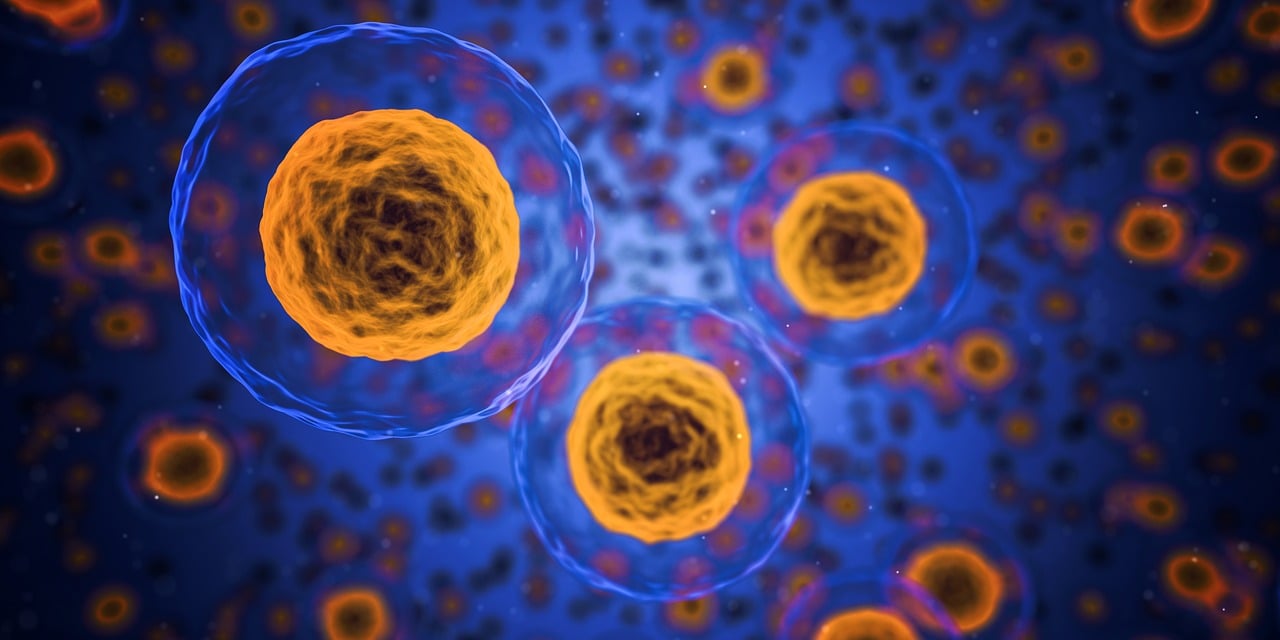
Communication is the key. Imagine a crowd of people speaking different languages. You are trying to convey an important message to the person standing on the other side of the room, but the communication paths leading to that person are like playing the broken telephone game – the information reaching the person at the end of the line is often completely different from what was originally intended.
Just like for us humans, proper communication is essential for the proper functioning of all the cells that make up our bodies. Throughout the embryonic development, during the response to a bacterial infection, or in the course of formation of new memories in the brain – cells pass the necessary information to each other. When this communication fails, it leads to abnormal development or to a disease.
How is the information transmitted in cells?
Cell communication is the main task governed by proteins called kinases. Kinases act as a courier service, delivering messages by attaching a phosphate group to their recipient proteins, molecules–“executors” in the cell. Acquisition of a phosphate group is like a message sent to a protein with instructions on what to do.
There are more than 500 different kinases in human cells, and most of them can transmit information to at least a few different recipient proteins. The number of possible communication paths is therefore really large, and the interpretation of communication extremely challenging due to the overlapping effects. Thus, cells are like crowded and full of communication paths cities, with lots of intersections and possible ways to get from the starting point to a remote part of the “city”. Hence, how to understand individual paths in this dense urban jungle?
In our laboratory, we have developed a technique that allows to direct the signal, channeling communication to the selected path in the cell. In other words, we can now decide which of the multiple potential recipient proteins send the information to. Our method can help understand better the single communication lanes that coordinate the development of an organism and its proper function, as well as interpret better the faulty communication in complex diseases such as cancer. It also opens up opportunities to create completely new communication paths in cell.
The principle of our approach is quite simple. We combined an activated catalytic domain of a chosen kinase (the part of the kinase that “does the job” of transferring information – the phosphate group) with a small antibody fragment, so called nanobody. The nanobody enables recognition of the selected recipient protein based on the added tag – here: GFP (Green Fluorescent Protein, see Fig. 1).
Thus, our engineered kinase is designed to preferentially activate the selected recipient protein, and not all proteins normally subject to a given kinase. And therefore – direct the information to the selected path in the complicated communication meshwork.
Our main protagonist is a fruit fly embryo (Drosophila melanogaster) – a great model to study animal development. Thanks to the genetic tools available in fruit fly, we can compare side by side cells in which the engineered kinase is produced and the unaltered, control cells.
Using advanced microscopy, we tested the effectiveness of our approach. We assessed the levels of phosphorylation (activation of a given protein by a kinase) and followed the development of tissues and organs in the developing fly embryo.
I will focus here on only one selected example. During the development of fly embryo into the larvae, an oval gap on the dorsal side of the embryo must be closed by stretching of the epithelial cells on both sides and “zippering” of the fissure. This phenomenon is important not only in flies – similar mechanisms control wound healing and neural tube closure during the fetal development. These processes involve the cytoskeleton (literally “cellular skeleton”), a network of filaments that forms the scaffold of the cell, giving it a shape and allowing the cell to move.
In our research, we used the described process of embryo closure as a model to verify the engineered kinases on the example of Rok kinase (ROCK in vertebrates). We proved high activity of the engineered Rok kinase by demonstrating that it robustly phosphorylates labeled myosin light chain protein. This further lead to the formation of large clusters of actomyosin (Photo 1), which is a component of the apparatus responsible for cell contractility and flexibility in the developing embryo.
In this way, we created and tested engineered kinases based on two different enzymes – Rok and Src kinase, linked to different nanobodies. We showed that these kinases efficiently transferred information to a chosen tagged protein, allowing for targeting cellular communication preferentially to a selected pathway, in a living organism.
We assume that after careful optimization our approach can be adapted to other kinases and recipient proteins in various cells and model organisms. The next step we are currently working on is to precisely control the time and place of “switching on” our kinase to transmit information by using light of the appropriate wavelength (for the initiated: combining our method with optogenetics).
Editorial Board’s Note: This discovery could significantly impact the medical sector, consequently affecting our daily lives.
Katarzyna Lepeta


